Science
Scientists Explore Human Anatomy Through Unique MRI Experiment
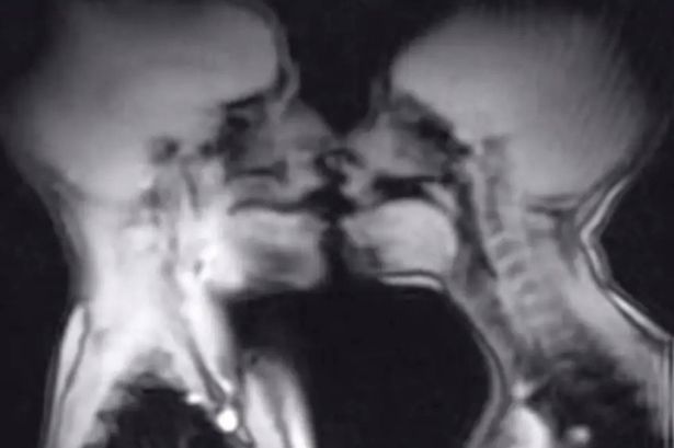
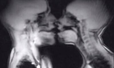
A couple’s intimate encounter inside an MRI scanner has provided scientists with intriguing insights into human anatomy. Ida Sabelis and her boyfriend Jupp participated in a study published in the British Medical Journal in 1999, aiming to understand physiological changes during sexual activity. The experiment revealed unexpected findings about both male and female anatomy during intercourse.
The research demonstrated that capturing magnetic resonance images of the male and female genitals during sex is feasible. It notably showed that during intercourse in the “missionary position,” the penis adopts a shape resembling a boomerang, with approximately one-third of its length comprising the root of the penis. Additionally, the study observed that during female sexual arousal without intercourse, the uterus elevates, and the anterior vaginal wall lengthens, although the size of the uterus itself does not increase during arousal.
Despite these findings, one aspect puzzled researchers. Across all 13 instances of sexual activity recorded in the MRI machine, it was noted that the women’s bladders filled rapidly. According to expert Menko Victor ‘Pek’ van Andel, this phenomenon may suggest an evolutionary mechanism intended to encourage women to urinate post-coitus, potentially reducing the risk of urinary tract infections.
In a further exploration of the experiment, Sabelis expressed that while the experience lacked romanticism, it served a greater purpose. She is an advocate for women’s rights and expressed a desire to enhance understanding of female physiology in scientific research. Describing the experience as both an “act of love” and a “performance,” Sabelis highlighted the importance of such studies in broadening the scope of women’s health.
While the MRI study was deemed a success, it also serves as a reminder of the importance of safety during medical procedures. In a separate incident, a woman sustained severe injuries after a metal-containing sex toy was inadvertently left inside her body during an MRI scan. The 22-year-old believed her “butt plug” was entirely made of silicone; however, its metal core reacted with the MRI machine, resulting in the dangerous migration of the toy through her body. Reports circulated online, warning others about the dangers of using metallic objects before MRI appointments.
The contrasting outcomes from these experiences emphasize the critical balance between scientific exploration and patient safety in medical practices. As research continues to evolve, studies like that of Sabelis and Jupp contribute valuable insights into understanding human anatomy, while also highlighting the need for stringent safety measures during medical imaging procedures.


Belfast Gears Up for Vibrant St Patrick’s Day Parade on March 17

Cuba Faces Nationwide Blackout Amid Deepening Energy Crisis

Wirral Council to Restructure Leadership, Cutting Two Top Roles

Rethinking Resistance Training: Key Insights from US Experts

EU Membership on the Horizon: What It Means for Citizens

Ukrainian Drone Swarms Strike Moscow Region for Second Day

Henley Launches Innovative Initiative to Boost Local Economy

Court Ruling Allows Pandemic Tax Refund Claims Until 2026

Sean Penn’s Absence at 2026 Oscars Sparks Discussion

Coronation Street’s Shocking Murder Twist Reveals Family Secrets

Andrew Pierce Confirms Departure from ITV’s Good Morning Britain

Katie Price Faces New Health Concerns After Cancer Symptoms Resurface

Gyles Brandreth Shares Grandson’s Cancer Battle and Recovery

Sue Radford Reveals Weight Loss Journey, Shedding 12–13 kg

Kate Garraway Sells £2 Million Home Amid Financial Struggles

Jordan Brook Faces Health Crisis in Hospital as Sophie Kasaei Stays Away

EastEnders’ Nicola Mitchell Faces Unexpected Pregnancy Crisis

Reddit Study Reveals Distinctions Between Lurkers and Power Users
-
 World8 months ago
Coronation Street’s Shocking Murder Twist Reveals Family Secrets
-
 Entertainment8 months ago
Andrew Pierce Confirms Departure from ITV’s Good Morning Britain
-
 Health11 months ago
Katie Price Faces New Health Concerns After Cancer Symptoms Resurface
-
 Health6 months ago
Gyles Brandreth Shares Grandson’s Cancer Battle and Recovery
-
 Health6 months ago
Sue Radford Reveals Weight Loss Journey, Shedding 12–13 kg
-
 Entertainment12 months ago
Kate Garraway Sells £2 Million Home Amid Financial Struggles
-
 Entertainment4 months ago
Jordan Brook Faces Health Crisis in Hospital as Sophie Kasaei Stays Away
-
 World9 months ago
EastEnders’ Nicola Mitchell Faces Unexpected Pregnancy Crisis
-
 Science8 months ago
Reddit Study Reveals Distinctions Between Lurkers and Power Users
-

 World8 months ago
Mother Charged After Son, 9, Killed in Tragic Incident in Italy
-

 Entertainment11 months ago
Ann Ming Reflects on ITV’s ‘I Fought the Law’ Drama
-

 World8 months ago
Bailey Announces Heartbreaking Split from Rebecca After Reunion