Science
New Quantum Biosensor Developed from Fluorescent Protein

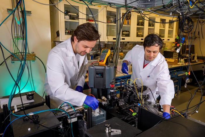
Researchers at the University of Chicago Pritzker School of Molecular Engineering have developed a groundbreaking quantum biosensor utilizing a new optically addressable quantum bit (qubit) encoded in a fluorescent protein. This innovative device can be produced directly within living cells, marking a significant advancement for fluorescence microscopy in monitoring biological processes.
Quantum technologies harness the unique properties of qubits to store and process information. Unlike classical bits, which can only exist in a state of either 0 or 1, qubits can be in a superposition of both states. This capability enables quantum computers to process multiple information streams simultaneously, allowing them to tackle complex problems far more efficiently than traditional computers. In sensing applications, qubits serve as nanoscale probes that can detect minute environmental changes with exceptional sensitivity.
Advancements in Quantum Sensing
Optically addressable qubit sensors can measure nanoscale magnetic fields, electric fields, and temperature. Currently, these sensors are widely used in the physical sciences, yet their application in life sciences remains limited, with most implementations still at the proof-of-concept stage. Existing quantum sensors often rely on nitrogen-vacancy (NV) centers, which are defects in diamond that behave as tiny quantum magnets.
According to Peter Maurer, co-lead of the study, the size of these sensors—typically ten times larger than most proteins—complicates their integration into living cells. Maurer explained, “The problem is that such sensors are difficult to position at well-defined sites inside living cells.” To overcome this challenge, the research team, led by Maurer and David Awschalom, opted to develop a qubit from a biological system itself.
The fluorescent proteins, measuring just 3 nm in diameter, can be genetically encoded, allowing cells to produce these sensors directly at desired locations. This unique capability has established fluorescent proteins as the “gold standard” in cell biology. Decades of research have created a vast library of these proteins, which can be tagged to various biological targets.
Maurer and Awschalom noted, “We recognized that these proteins possess optical and spin properties that are strikingly similar to those of qubits formed by crystallographic defects in diamond.” They combined fluorescence microscopy techniques with quantum control methods to encode and manipulate protein-based qubits.
In their study, published in Nature, the researchers employed a near-infrared laser pulse to optically address a yellow fluorescent protein known as EYFP. They achieved a triplet spin state readout with impressive results, showing up to 20% “spin contrast” measured using optically detected magnetic resonance (ODMR) spectroscopy.
Potential Applications in Biological Research
To evaluate the technique’s effectiveness, the team genetically modified the EYFP protein for expression in human embryonic kidney cells and Escherichia coli cells. The measured ODMR signals demonstrated a contrast of up to 8%. While this performance does not yet match that of NV quantum sensors, the fluorescent proteins present a new opportunity for magnetic resonance measurements directly within living cells—something NV centers cannot achieve.
Maurer emphasized the transformative potential of this technology, stating, “They could thus transform medical and biochemical studies by probing protein folding, monitoring redox states, or detecting drug binding at the molecular scale.”
Additionally, the quantum resonance signatures from these sensors offer a new dimension for fluorescence microscopy, paving the way for highly multiplexed imaging beyond current capabilities. Looking to the future, the researchers are exploring the use of arrays of such protein qubits to investigate many-body quantum effects within biologically assembled structures.
The research team is currently focused on enhancing the stability and sensitivity of their protein-based qubits through protein engineering techniques akin to the “directed evolution” used to optimize fluorescent proteins for microscopy. They aim to achieve single-molecule detection, enabling the readout of the quantum state of individual protein qubits within cells.
Their ongoing efforts also include expanding the range of available qubits by investigating new fluorescent proteins with improved spin properties and developing sensing protocols capable of detecting nuclear magnetic resonance signals from nearby biomolecules. This could reveal structural changes and biochemical modifications at the nanoscale, opening new avenues for scientific inquiry and discovery.
-

 World2 months ago
Coronation Street’s Shocking Murder Twist Reveals Family Secrets
-

 Entertainment2 months ago
Andrew Pierce Confirms Departure from ITV’s Good Morning Britain
-

 Health6 months ago
Katie Price Faces New Health Concerns After Cancer Symptoms Resurface
-

 Health4 weeks ago
Sue Radford Reveals Weight Loss Journey, Shedding 12–13 kg
-

 Entertainment7 months ago
Kate Garraway Sells £2 Million Home Amid Financial Struggles
-

 World3 months ago
EastEnders’ Nicola Mitchell Faces Unexpected Pregnancy Crisis
-

 Entertainment6 months ago
Ann Ming Reflects on ITV’s ‘I Fought the Law’ Drama
-

 World3 months ago
Bailey Announces Heartbreaking Split from Rebecca After Reunion
-

 Entertainment3 months ago
Coronation Street Fans React as Todd Faces Heartbreaking Choice
-

 Entertainment2 months ago
David Jason and Nicholas Lyndhurst Eye Reunion for Only Fools Anniversary
-

 Entertainment3 months ago
Bradley Walsh Sparks Strictly Come Dancing Hosting Speculation
-

 Entertainment2 months ago
Two Stars Evicted from I’m A Celebrity Just Days Before Finale


